- Libreria
- >
- Libri in lingua
- >
- Medicina
- >
- Istologia
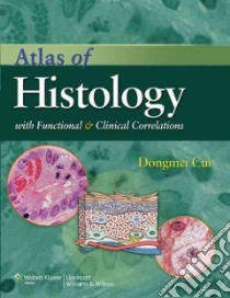
Atlas of Histology - 9780781797597
Un libro in lingua di Dongmei Cui Naftel John P. Ph.D. Daley William P. M.D. Lynch James C. Haines Duane E. edito da Lippincott Williams & Wilkins, 2010
- € 81.10
- Il prezzo è variabile in funzione del cambio della valuta d’origine
With the first section containing an illustrated glossary of histological and pathological terms, this color atlas is organized into three sections, progressing from cell structure and function through basic tissues and 13 complex organ systems (including the oral cavity, the eye, and the ear). About 1,300 illustrations and micrographs, including color and b&w histology slides at low- and high-power magnifications, high-quality anatomic and process illustrations, and a few color clinical photos, all fully labeled with complete names to allow easy identification are provided. To demonstrate how tissues are modified by a pathological process, each page layout shows light and electron micrograph images and diagrams of normal and abnormal tissues side-by-side. In addition to images, the atlas offers explanatory text in each chapter and expanded figure captions, plus key concept summaries, clinical content boxes, and synopsis boxes on key structural and functional characteristics of cells, tissues, and organs. The book includes chapter summary tables, plus an appendix explaining the general concept of tissue preparation and staining. An online component provides searchable text and all images, plus an interactive labeling atlas. Cui teaches medical and dental histology at the University of Mississippi Medical Center. Annotation ©2011 Book News, Inc., Portland, OR (booknews.com)
Informazioni bibliografiche
- Titolo del Libro in lingua: Atlas of Histology
- Sottotitolo: With Functional and Clinical Correlations
- Autori : Dongmei Cui Naftel John P. Ph.D. Daley William P. M.D. Lynch James C. Haines Duane E.
- Editore: Lippincott Williams & Wilkins
- Collana: Lippincott Williams & Wilkins (Paperback)
- Data di Pubblicazione: 26 Luglio '10
- Genere: MEDICAL
- Argomenti : Histology Atlases Histology, Pathological Atlases Histology Atlases
- ISBN-10: 0781797594
- EAN-13: 9780781797597


